

Inside a vein or artery, these gel-like masses can block circulation even though they normally stop bleeding after an injury. A clot in the leg may cause swelling or pain, while a clot affecting the lungs, heart, or brain can become an emergency within minutes.
You can use the details below to recognize warning signs, understand clotting risk factors, and know when medical testing or emergency care is needed.
Normal Clotting Turns Dangerous Inside a Blood Vessel
After a cut, platelets collect at the damaged area and fibrin creates a mesh that stabilizes the plug. That process limits blood loss. Trouble begins when clotting occurs inside an intact vessel, where the blockage can reduce oxygen delivery to tissue downstream.
Your body also uses fibrinolysis, a cleanup process in which plasmin breaks down fibrin after healing starts. A harmful clot can overwhelm that system or form where blood moves too slowly. Location matters because venous and arterial blockages create different symptoms and require different levels of urgency.
| Clot category | Where it forms | What can happen |
|---|---|---|
| Venous thrombosis | Deep veins, commonly in a calf or thigh | Part of the clot can travel through the bloodstream to the lungs. |
| Deep vein thrombosis, or DVT | Deep leg, pelvic, or arm vein | Swelling and tenderness may appear, although some cases cause no obvious sign. |
| Pulmonary embolism, or PE | Lung artery after travel from a vein | Oxygen exchange and heart function can become strained quickly. |
| Arterial blood clot | Artery feeding the heart, brain, or limb | A heart attack, stroke, or threatened limb can result. |
Venous thromboembolism, or VTE, is the umbrella term for DVT and PE. Arterial blood clots usually form through a different process: fatty plaque ruptures, platelets stick to the injured surface, and blood flow falls beyond the blockage. Those pathways explain why your next step depends on the symptom pattern and body area involved.
Because blocked vessels affect different tissues, warning signs can look very different from one body area to another.
Symptoms Change With the Clot Location
Because location changes the danger, new calf swelling and sudden breathlessness should not be treated as the same problem. A clot in a deep leg vein can break loose and reach the lungs, so a symptom that starts in one place can become urgent somewhere else.
Leg, Lung, Heart, Brain, and Limb Symptoms Need Different Responses
A one-sided swollen calf needs prompt evaluation, while sudden chest pain or a face droop requires emergency action. You cannot confirm a vascular blockage by appearance alone, but the pattern and speed of symptoms help determine where to seek care.
| Possible location | Signs that may appear | Action |
|---|---|---|
| Leg or arm DVT | New one-sided swelling, warmth, pain, tenderness, redness, or darker skin tone | Seek clinical care that day. |
| Lung PE | Sudden shortness of breath, chest pain, rapid heartbeat, fainting, or coughing blood | Call 911. |
| Heart artery | Chest pressure, sweating, nausea, breathlessness, or pain spreading to an arm, jaw, back, or shoulder | Call 911. |
| Brain artery | Face droop, arm weakness, speech trouble, sudden confusion, vision change, or severe balance loss | Call 911. |
| Abdomen or limb artery | Severe belly pain, a cold pale limb, numbness, or severe pain out of proportion to appearance | Seek emergency medical care. |
A muscle cramp may ease with rest, whereas DVT discomfort can persist with swelling, skin color change, or heat. Your leg can look nearly normal despite a deep clot, so no redness does not rule out a problem. These blood clot symptoms deserve same-day care when they are new, one-sided, or unexplained.
Emergency Symptoms Need an Ambulance Response
Trouble breathing, fainting, slurred speech, or sudden circulation changes require emergency assessment rather than home monitoring. Call 911 instead of driving yourself because emergency crews can begin evaluation during transport.
- Sudden breathing trouble: Call 911 for new unexplained breathlessness, especially with chest pain or a racing pulse.
- Chest pressure: Treat squeezing, heavy, or sharp chest discomfort as an emergency until clinicians rule out a heart or lung cause.
- Fainting or collapse: Loss of consciousness can signal reduced blood flow and needs an ambulance response.
- Stroke warning signs: Face drooping, speech changes, or one-sided weakness require 911 right away.
- Severe limb distress: A suddenly cold, blue, numb, or intensely painful arm or leg needs emergency evaluation.
Call 911 rather than driving yourself when PE, stroke, heart attack, fainting, or severe circulation symptoms are possible. Emergency crews can begin assessment during transport.
New one-sided limb swelling or pain without breathing or neurologic symptoms also warrants clinical care that day. Your personal risk profile helps clinicians decide how urgently to test, which leads to the causes behind abnormal clotting.
Behind those varied symptoms, several conditions can make blood more likely to clot when it should not.
Slow Flow, Vessel Injury, and Hypercoagulability Raise Risk
Those symptom patterns become more concerning when one or more major clotting conditions are present. Clinicians use Virchow’s triad to describe three forces behind thrombosis: sluggish flow, vessel-wall injury, and hypercoagulability, meaning blood has a stronger tendency to form a clot.
A long hospital stay can involve all three parts of Virchow’s triad at once. Surgery injures tissue, bed rest slows blood return from the legs, and serious illness can make clotting activity more intense. These blood clot causes and risk factors can overlap rather than appear one at a time.
Several Risk Factors Can Stack Together
Risk rises through combinations, not just a single diagnosis or event. Your clinician will usually ask about recent movement, operations, medicines, pregnancy, cancer history, and any earlier DVT or PE.
- Limited movement: Bed rest, a long flight, or hours seated after an injury can slow venous return from the legs.
- Recent surgery: Tissue injury and reduced mobility can raise risk during recovery, particularly after major operations.
- Serious illness: Cancer, severe infection, and hospitalization can shift the body toward more active clotting.
- Prior event: A previous DVT or PE changes future risk and follow-up needs.
- Tobacco exposure: Smoking damages blood vessels and adds cardiovascular risk that can compound other factors.
- Higher body weight: Obesity can contribute through inflammation, reduced mobility, and added pressure on leg veins.
Pregnancy and the weeks after delivery change circulation and clotting proteins, making postpartum planning especially important. Estrogen-containing birth control and hormone therapy can also affect risk. A clinician may ask whether relatives have Factor V Leiden or a prothrombin gene change, two inherited traits linked with greater clotting tendency.
Risk factors do not diagnose a clot, and some events occur without a clear trigger. You may have several risks and never develop VTE, while another person develops symptoms after a short period of immobility. That uncertainty makes medical testing more reliable than self-diagnosis.
Since risk factors cannot confirm a clot, clinicians rely on targeted questions, tests, and scans to clarify the cause.
Diagnosis Combines History, Lab Results, and Imaging
Because risk alone cannot confirm the problem, clinicians match testing to your symptoms and likelihood of disease. A sore calf can resemble a strain, and chest tightness can overlap with anxiety, so home checks cannot safely settle the question.
A clinician reviews symptom timing, body location, medicines, recent travel, surgery, prior health history, and physical findings. Your pregnancy status, kidney function, contrast allergy, and symptom severity can also affect which scan is safest and most useful.
Testing Depends on the Suspected Site
No single test fits every situation. Lower-risk cases may start with a blood test, while severe chest, neurologic, or limb symptoms can require imaging without delay.
- Risk review: Clinical scoring tools help sort low, intermediate, and higher suspicion before imaging is chosen.
- D-dimer test: This blood test detects fibrin breakdown fragments and can help rule out VTE in selected lower-risk settings.
- Ultrasound imaging: Compression ultrasound checks whether a leg vein collapses normally or contains material blocking the lumen.
- Lung imaging: CT pulmonary angiography can show blocked lung arteries, while a ventilation-perfusion scan may suit selected situations.
- Urgent organ scans: ECGs, heart imaging, CT, or MRI can show signs linked to a suspected heart attack or stroke.
The D-dimer test cannot identify a clot by itself because surgery, pregnancy, infection, and aging can raise the result. Ultrasound imaging uses sound waves rather than radiation, making it a central test for suspected DVT. That approach matches Centers for Disease Control and Prevention guidance describing VTE as a serious condition that needs timely medical care.
Imaging identifies the location, while the exam shows how stable your circulation is. Those findings determine whether treatment can happen at home, needs close monitoring, or requires emergency hospital care.
Once severity and location are clear, care focuses on preventing extension while protecting vital circulation.
Medical Care Stops Growth and Reduces New Clot Formation
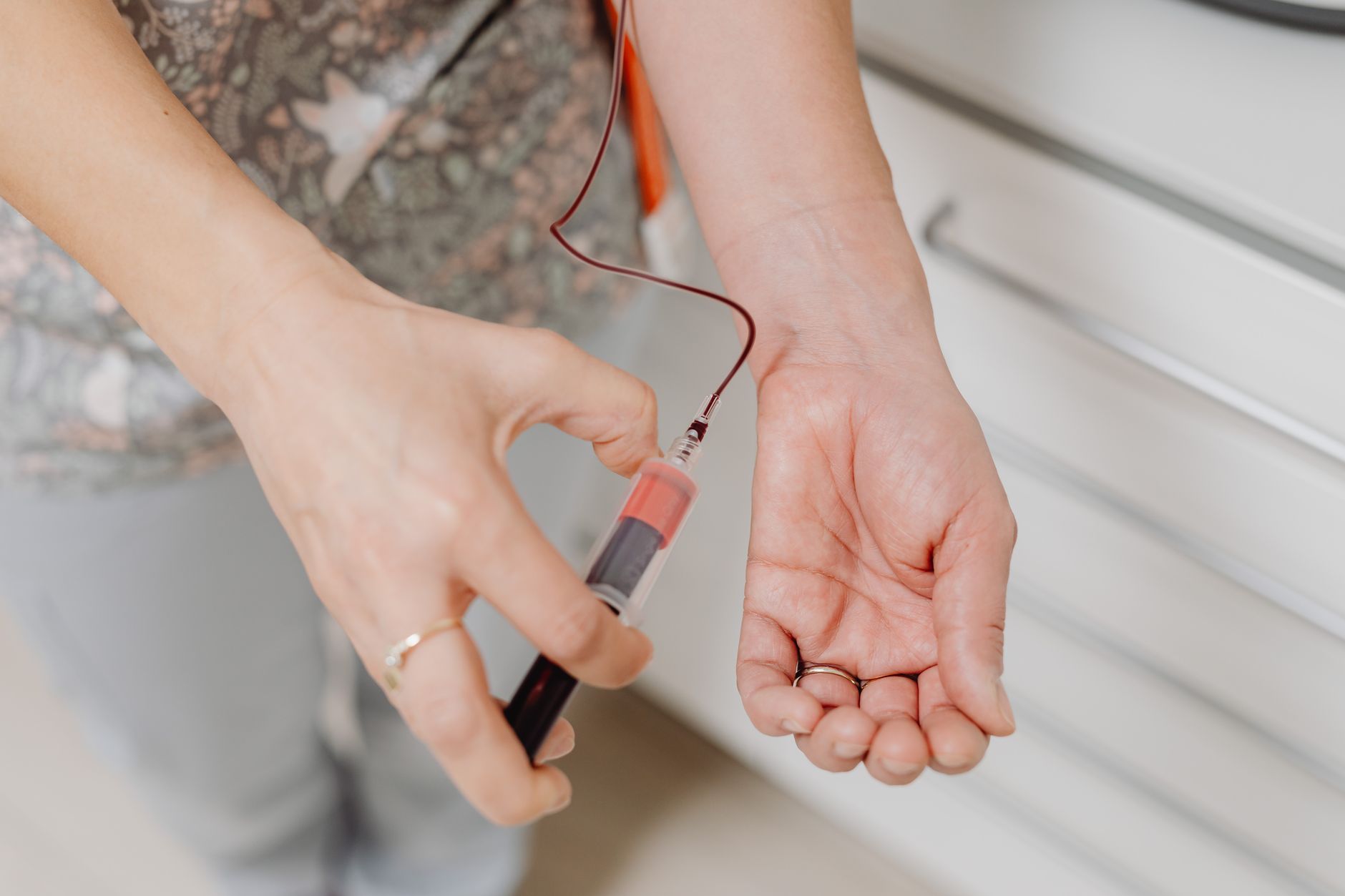
Once testing identifies the location and severity, treatment aims to prevent enlargement and dangerous movement. Anticoagulants, often called blood thinners, are the main blood clot treatment for many DVTs and PEs.
Despite the name, an anticoagulant does not thin blood like water or directly dissolve most existing clots. It slows the body’s ability to make new clot material while natural fibrinolysis clears the blockage over time. Your treatment duration can differ after surgery, during cancer care, in pregnancy, after an unexplained event, or after recurrence.
Severe Events May Need Faster Procedures
A severe PE, acute ischemic stroke, heart attack, or limb with failing circulation may require faster treatment than anticoagulants alone. Specialists use imaging, blood pressure, oxygen levels, and bleeding risk to decide whether clot-busting medication, a catheter procedure, surgery, or hospital intervention is appropriate.
Restoring blood flow quickly matters because heart muscle, brain tissue, and limb tissue can sustain lasting injury without oxygen. Your care team weighs that benefit against bleeding risk before using more aggressive treatment. That clinical balance aligns with American Heart Association information on rapid treatment in heart and brain emergencies.
Medication Safety Prevents Avoidable Bleeding
Anticoagulants work best when taken on schedule and reviewed alongside every other medicine. Your prescriber or pharmacist needs to know about new prescriptions, supplements, and over-the-counter pain medicines.
- Follow the label: Take anticoagulant medication exactly as prescribed and contact the prescriber before any change.
- Report unusual bleeding: Black stools, vomiting blood, heavy unexplained bleeding, or a serious fall need urgent medical advice.
- Check new products: Ask a pharmacist or clinician before starting over-the-counter drugs, supplements, or NSAID pain relievers.
- Share your medication list: Tell dentists, surgeons, and urgent-care staff about anticoagulant use before procedures.
Do not rely on supplements, massage, or home remedies to dissolve a suspected vascular clot. Massage over a painful swollen limb can delay the evaluation your situation needs.
Medication interactions can increase bleeding risk even when the anticoagulant dose is correct. Food and Drug Administration medication safety materials support checking other drugs before combining them with anticoagulants. Prevention planning becomes especially useful after the immediate danger has passed.
After treatment begins, everyday habits and advance planning can help lower the chance of another event.
Movement and Planning Reduce Preventable Risk
After treatment decisions are clear, daily movement can support better blood flow in the legs. Calf muscles act like a pump for venous return, so standing, walking, and ankle flexing interrupt long stretches of stillness.
During desk work or travel, change position and walk at intervals rather than remaining seated for several hours. Hydration may fit your health plan, but fluid limits can apply with heart or kidney conditions. Blood clot prevention works best when it is tailored to your surgery, travel, mobility, and medical history.
Different Settings Need Different Plans
The right plan depends on why risk is elevated and how long that period will last. Your discharge instructions and clinical history matter more than a generic travel or recovery checklist.
- After surgery: Follow discharge instructions about walking, compression measures, and any prescribed anticoagulant plan.
- During travel: Stand, stretch, and walk during longer trips whenever mobility and the travel setting allow.
- After illness: Ask about movement goals before leaving the hospital, especially after a prolonged bed stay.
- For recurrence: Keep follow-up visits and discuss new symptoms, missed doses, smoking cessation, or planned procedures.
- Before hormones: Review estrogen options with a clinician when a history includes VTE or strong family risk.
- For pregnancy planning: Bring prior clot history to obstetric care before or soon after pregnancy begins.
Compression stockings and preventive anticoagulants may be part of surgical planning, but neither is right for every person. The surgical team considers bleeding risk, procedure type, mobility, and past history before setting that plan. Your safest approach is to follow the prevention plan created for the specific procedure or health condition.
Menstrual clots are shed uterine lining and blood during a period, not a clot inside a vein or artery. Large or frequent menstrual clots deserve gynecologic assessment, yet they do not by themselves point to DVT or PE. New limb swelling, chest pain, breathlessness, fainting, stroke signs, or severe circulation changes need medical care without delay.
Even with sensible precautions, recognizing urgent warning signs remains the most important takeaway.
Bottom Line
A normal clot seals an injury, while a vessel blockage can interrupt blood flow where organs need it most. Your response should match the symptom pattern and speed of onset: arrange same-day clinical care for new one-sided swelling or pain, and call 911 for sudden breathing trouble, chest symptoms, fainting, stroke signs, or severe limb pain.
Fast evaluation can identify DVT, PE, arterial blockage, or another cause of your symptoms. The details that matter most are where symptoms began, how quickly they changed, and whether recent illness, travel, surgery, pregnancy, or immobility raised your risk.
FAQ
What are the first signs of a blood clot?
Early signs of a DVT can include new swelling on one side, calf or arm tenderness, warmth, redness, or skin discoloration. Some deep vein clots produce few visible signs, so persistent new symptoms deserve same-day clinical assessment, particularly after travel, surgery, illness, or reduced movement.
What are the first signs of a blood clot in the leg, arm, lung, heart, or brain?
Signs of a blood clot in a leg or arm can include one-sided swelling, pain, warmth, tenderness, or color change. A lung clot can cause sudden breathlessness, chest pain, fainting, or coughing blood. Heart and brain symptoms can include chest pressure, sweating, face drooping, speech trouble, sudden weakness, or confusion, and each needs a 911 call.
What usually causes blood clots?
Clots form more readily when blood flow slows, a vessel is injured, or clotting tendency rises. Surgery, injury, hospitalization, immobility, cancer, infection, smoking, obesity, pregnancy, estrogen exposure, inherited clotting disorders, and a prior event can all add to your risk profile.
What is the difference between DVT, pulmonary embolism, and an arterial clot?
DVT is a clot in a deep vein, usually in a leg, pelvis, or arm. A pulmonary embolism occurs when clot material reaches and blocks an artery in the lungs. An arterial clot blocks an artery supplying the heart, brain, or limb and can cause a heart attack, stroke, or threatened circulation.
Are blood clots very serious?
They can be serious because a clot may block blood supply in a leg, lung, heart, brain, or other organ. A pulmonary embolism, heart attack, stroke, or sudden threatened limb circulation needs emergency care, while suspected DVT also needs prompt clinical evaluation.
Will blood clots go away on their own?
Your body can gradually break down clot material, but waiting without medical assessment can be dangerous because a clot may grow, travel, or block circulation. A clinician can identify the site, judge the risk, and set care based on imaging, symptoms, and your medical history.
