

Demodectic Mange is a skin disease caused by an excessive number of host-specific Demodex mites in your dog’s hair follicles. It can start as a few subtle bald spots around the eyes or muzzle, then range from a mild juvenile concern to a sign of deeper illness that needs veterinary investigation.
You’ll find the practical details on mite biology, visible skin changes, diagnosis, treatment, recovery checks, and the warning signs that call for prompt veterinary care.
Follicle Mites Drive Demodicosis
Most dogs carry small numbers of microscopic Demodex mites without visible skin trouble. Demodectic mange develops when the mite population grows beyond what the skin and immune defenses can control.
Canine Demodex Mites Stay Within Their Host Species
Demodex canis causes many canine cases. Its long, cigar-shaped body fits inside hair follicles, where it feeds on cellular material and moves through egg, larval, nymph, and adult stages.
Another species, Demodex injai, has a longer body and may be linked with greasy or oily skin changes in some dogs. Your veterinarian can identify these mites under a microscope because their shape and follicle location differ from other skin parasites.
Dogs usually acquire Demodex mites from their mother during the first days of life through close skin contact. That normal transfer does not create a household outbreak because canine Demodex mites are adapted to dogs, not people, cats, or healthy adult dogs.
Immune Control Limits Mite Numbers
Inside hair follicles, T-cell immune responses help keep mite populations low. Puppies have maturing immune regulation, while some dogs may have inherited tendencies that make follicle control less reliable during juvenile-onset demodicosis.
Skin damage, poor nutrition, hormonal disease, stress, and immune-suppressing medicines can also change that balance. Your veterinarian looks beyond the mites because sudden widespread disease in a mature dog can point to a separate health problem.
Demodex mites are not considered a routine contagion threat to your family or your other pets. New bald patches still need veterinary attention because several contagious skin diseases can look similar.
That host-specific biology removes one common worry, but the pattern of hair loss still matters. Allergies, fungal disease, flea reactions, and sarcoptic mites can all resemble follicular mite disease, so the next step is to examine the skin changes closely.
Because mimicry is common, the arrangement and progression of hair loss become especially useful diagnostic clues.
Hair Loss Patterns Offer Early Clues
Because household spread is rarely the main concern, lesion shape and location provide useful clues. Small circles of missing coat around the eyes, lips, muzzle, face, and front legs are familiar early demodectic mange symptoms.
Mild Lesions Can Look Like Small Bare Spots
A patch may begin as a thin area no larger than a quarter, with flakes trapped in the remaining fur. Redness, small bumps, blackheads, and dry scale can follow as inflamed follicles lose hair.
Your dog may scratch very little at this stage. Mild or absent itching does not rule out demodicosis because the mites themselves may cause less itch than allergic skin disease or sarcoptic mange.
Advanced lesions can include thickened skin, darker pigmentation, crusting, and a stale or musty odor. Strong odor and sudden intense scratching raise concern for a secondary bacterial skin infection rather than mite activity alone.
Several Skin Conditions Share Similar Surface Signs
| Condition | Pattern you may see | Clue that helps separate it |
|---|---|---|
| Demodicosis | Patchy hair loss, scale, redness, facial or leg lesions | Deep skin scraping may reveal mites within follicles |
| Sarcoptic mange | Marked itching, ear-edge crusts, belly and elbow irritation | Close-contact spread is a greater concern for dogs and people |
| Ringworm | Round hairless areas with scale or broken hairs | Fungal culture or PCR helps identify dermatophyte infection |
| Flea allergy dermatitis | Chewing near the tail base, rump, and rear thighs | Flea dirt or flea exposure supports the pattern |
| Bacterial folliculitis | Pustules, crusts, circular collars of scale | Skin cytology may show bacteria and inflammatory cells |
| Allergic skin disease | Feet, ears, belly, and recurrent itch | Seasonal flares or environmental exposure may fit your dog’s history |
For demodectic mange vs sarcoptic mange, itch level and contagion risk create the practical dividing line. Sarcoptic disease becomes more likely in a wildly itchy dog with affected housemates, while follicular patches with limited scratching fit Demodex more closely, though testing settles the diagnosis.
Surface appearance cannot show how far the disease has spread below the coat. Distribution across your dog’s body changes the urgency, prognosis, and veterinary plan.
Yet a recognizable pattern reveals little about whether the problem remains localized or has become medically serious.
Extent of Skin Disease Changes the Medical Stakes
A few isolated patches in a young dog have a different outlook than painful, inflamed skin across the trunk, feet, and face. Localized vs generalized demodicosis describes disease extent, not merely how dramatic the coat looks.
Localized Demodicosis Stays Limited to Small Areas
Veterinarians use localized demodicosis for a small number of affected areas confined to one part of the body. A young dog with two coin-sized bald spots near the eyes and no paw involvement may fall into this category.
Some juvenile localized cases recede as immune regulation matures, so observation rather than immediate mite-directed medication may be appropriate. Your dog still needs rechecks because a rising patch count or new body regions change that decision.
Generalized Demodicosis Requires Closer Care
Generalized demodicosis affects many sites, large body regions, or an entire area such as the face, legs, trunk, or paws. Pododemodicosis, which affects the feet, deserves special attention because deep swelling and infection can make it difficult to resolve.
- Painful sores: Open, tender skin can signal deeper inflammation or bacterial involvement that needs prompt veterinary care.
- Pustules and drainage: Yellow material, crusts, or wet lesions may indicate pyoderma inside damaged follicles.
- Foot swelling: Enlarged paws, limping, or pain between the toes can accompany difficult pododemodicosis.
- Foul skin odor: A sharp smell suggests bacterial overgrowth or heavy skin debris rather than a mild hairless patch.
- Whole-body changes: Fever, low energy, poor appetite, or broad hair loss warrant a same-day veterinary call.
Severe generalized disease complicated by deep pyoderma can make a dog seriously ill, although mites alone do not usually cause fatal illness. The greater danger comes from damaged skin, bacterial invasion, pain, fever, and systemic illness.
Do not wait on pustules, fever, lethargy, bleeding skin, or rapidly spreading hair loss. These signs need veterinary assessment rather than a home shampoo experiment.
The difference between mild and severe disease cannot rest on photographs alone. Finding mites and identifying bacterial or yeast complications is where veterinary diagnosis becomes essential.
That uncertainty makes laboratory confirmation crucial, particularly when secondary infections can alter both appearance and risk.
Veterinary Testing Separates Demodicosis From Look-Alikes
Microscopy provides the clearest evidence because Demodex mites live below the skin surface. A veterinarian may squeeze the skin to push follicle contents upward, then collect a deep skin scraping until mild capillary bleeding appears.
Different Samples Answer Different Skin Questions
Deep skin scrapings are a core method for finding Demodex canis eggs, larvae, nymphs, and adults. Hair plucks, also called trichograms, can collect follicular material from delicate areas such as around the eyes.
| Veterinary method | What it samples | Why it may be selected |
|---|---|---|
| Deep skin scraping | Follicle contents and superficial skin debris | Useful for finding and counting mites in active lesions |
| Hair pluck | Hairs and material attached to roots | Helpful near thin skin or sensitive facial areas |
| Tape preparation | Surface cells, yeast, bacteria, and debris | Helps identify infection alongside mite disease |
| Skin cytology | Inflammatory cells and microorganisms | Guides care for pustules, odor, and moist lesions |
| Skin biopsy | Deeper tissue structure | May help with thickened skin, feet, or repeated negative scrapings |
A negative scraping does not always end the search. Thick scar-like skin, infected paws, and deeply altered follicles can hold mites beyond the sample, so your veterinarian may collect several samples or use a biopsy.
Adult-Onset Disease Calls for a Broader Workup
A seven-year-old dog that develops bald patches across the neck, chest, and paws after years of normal skin needs investigation beyond mite control. Endocrine disorders, cancer, immune system dysfunction, nutritional problems, and medicines that suppress immunity can all reduce follicle defense.
Host immunity influences mite proliferation, which is why adult-onset demodicosis deserves more than a skin-only approach. That reasoning aligns with the Merck Veterinary Manual, which identifies immune status as a major factor in demodicosis.
Blood work, a medication review, and a history of weight change, thirst, appetite, or repeated infections may uncover a contributing illness. Once your veterinarian separates mites, bacteria, yeast, and look-alikes, treatment can target the actual problem instead of chasing a rash with random products.
With the cause clarified, care can address the parasites, complications, and health factors sustaining the skin damage.
Treatment Addresses Mites, Infection, and Underlying Disease
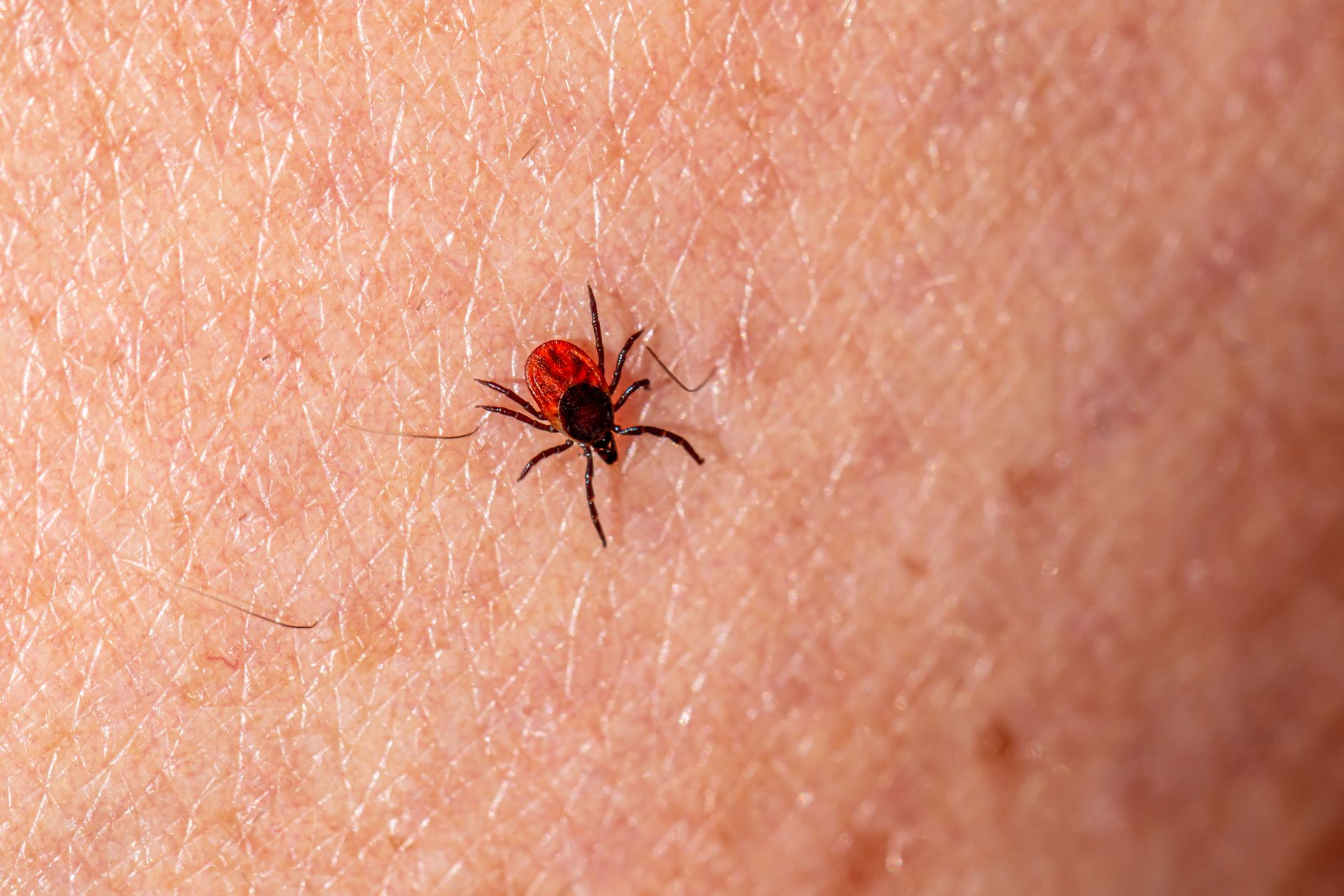
Care works best when it targets three problems at once: mite numbers, skin infection, and the factor that allowed follicular populations to increase. A single bath cannot manage all three, especially in a dog with generalized skin disease.
Modern Mite Control Uses Veterinary-Guided Medicines
Isoxazoline antiparasitic medications are oral parasite-control products that affect ligand-gated chloride channels in arthropods. Mammals use different receptor systems, which partly explains why these medications can act against mites, though your veterinarian still weighs age, breed, neurologic history, and concurrent illness.
Bravecto, NexGard, and Simparica are brand names associated with isoxazoline products. Labels, dosing schedules, and veterinary judgment differ, so your demodectic mange treatment plan should come from the clinician who examined the skin and reviewed the medical history.
Selected cases may need other acaricidal therapies alongside an isoxazoline plan. Herd products, concentrated farm formulations, and unverified internet remedies are poor substitutes because they may be harmful, unsuitable for dogs, or designed for parasites other than follicle mites.
Pyoderma Needs Separate Treatment
A secondary bacterial skin infection, also called pyoderma, can cause pain, pus, odor, fever, and deeper tissue damage. Veterinary care may include prescription antibiotics, medicated bathing, pain control, clip-and-clean care for dense coats, or other measures based on cytology and lesion depth.
- Bring clear photos: Photograph lesions every 7 days in the same light to show spread, redness, and coat return.
- List every product: Share collars, flea products, shampoos, supplements, and prescriptions so missed interactions are less likely.
- Limit skin picking: Reduce licking and chewing with veterinary guidance because self-trauma can push bacteria deeper into skin.
- Follow exact directions: Use the product label and your veterinarian’s instructions rather than doubling doses after a missed application.
- Keep visits scheduled: Recheck appointments reveal mite counts and infection changes that returning fur can hide.
Your dog may look more comfortable before the mite burden has dropped enough to stop treatment. That gap between visible improvement and microscopic evidence explains why recovery depends on repeat checks.
Visible regrowth is encouraging, but it cannot confirm that the underlying infestation has fully resolved.
Recovery Requires Follow-Up Beyond New Hair Growth
New fur can be misleading because follicles may produce hair while mites or bacterial inflammation remain active below the surface. That pattern is especially common after a widespread flare involving the trunk, legs, or paws.
Skin Comfort Improves Before Mite Clearance
Skin comfort may improve over several weeks, while a full coat can take longer to fill in. Darkened skin may also remain after inflammation settles because pigment changes fade more slowly than redness.
Your veterinarian may repeat skin scrapings or hair-based samples at planned intervals. Repeated negative mite findings, considered alongside the skin exam, help determine when mite-directed medication can stop.
Follow-Up Records Make Future Flares Easier to Judge
- Track lesion maps: Mark body sites on a simple dog outline so your veterinarian can compare new areas with recovering ones.
- Record skin changes: Write down odor, scratching, sleep disruption, appetite, and paw pain between appointments.
- Attend repeat checks: Bring your dog back on schedule even after fur returns because microscopic counts guide the next decision.
- Review prevention plans: Discuss year-round parasite control where your veterinarian advises it for your dog’s exposure and history.
- Discuss breeding carefully: Dogs with generalized juvenile-onset disease may have an inherited susceptibility worth addressing before breeding.
Inherited immune susceptibility may contribute to juvenile generalized disease, which affects breeding decisions. That position aligns with American Kennel Club advice against breeding dogs with generalized demodicosis.
Recurrence carries different meaning at different ages. A puppy with one returning patch and an older dog with new generalized lesions do not need the same medical search.
Age and species add important context, shaping whether similar-looking lesions warrant observation or a broader medical workup.
Cats and Mature Dogs Need Individualized Assessment
Age and species change the investigation because feline mites behave differently from canine mites. Demodectic mange in cats is less common than canine disease, and cat-specific diagnosis matters before treatment begins.
Feline Demodex Species Need Cat-Specific Care
Demodex cati lives deeper in cat hair follicles, while Demodex gatoi is shorter and lives closer to the skin surface. D. gatoi may spread between cats, unlike the usual canine Demodex pattern, and it can cause marked grooming, scale, and hair loss.
Your cat needs species-specific veterinary diagnosis before any parasite product is used. Products made for dogs may be unsafe for cats, and excessive grooming can also reflect allergy, pain, fungal disease, or another dermatologic disorder.
Mature Dogs Need More Than Mite Control
New generalized disease after maturity deserves a broader health assessment. Endocrine disease, cancer, long-term immune-suppressing drugs, and chronic systemic illness can weaken the control that once kept mites at low levels.
Bring a complete timeline of appetite, water intake, weight, medications, prior infections, and skin history. Details such as increased drinking or repeated urinary infections may lead your veterinarian toward a separate condition that needs treatment.
Routine household care remains sensible while the diagnosis is being clarified. Wash bedding and grooming tools, avoid sharing brushes during an unexplained rash, and isolate pets only where your veterinarian suspects a contagious alternative such as sarcoptic mange or ringworm.
Do not medicate other pets solely because one dog has follicular mites. Their skin signs, species, age, and parasite risk may be entirely different from your affected dog’s situation.
Even within one household, those differences underscore why each animal’s signs and risks deserve separate consideration.
Final Thoughts on Demodectic Mange
Patchy coat loss deserves a close look, but it does not automatically mean your home has a contagious infestation. Your strongest next step is a veterinary exam that identifies mites, checks for infection, and investigates deeper illness in adult-onset or widespread cases.
Careful follow-up matters as much as the first treatment choice. Decisions based on skin scrapings, lesion changes, and your dog’s overall health provide firmer answers than fur growth alone.
FAQ
These answers address the most common concerns about diagnosis, spread, treatment, and recovery. Your veterinarian should assess any dog with spreading bald patches, foot swelling, pustules, foul odor, fever, or low energy.
What is demodectic mange in dogs, and what causes it?
it in dogs is a skin disease caused by excessive numbers of host-specific Demodex mites in hair follicles. Immature immune control, inherited susceptibility, immune-suppressing medicines, endocrine disease, cancer, stress, or other immune system dysfunction can allow mite numbers to increase.
What are the first signs of demodectic mange in dogs?
Small bald patches around the eyes, muzzle, face, and front legs are common early signs. Your dog may also have dry scale, mild redness, or broken hairs, while itching may be absent or mild until infection or another skin problem develops.
Is demodectic mange contagious to people or other pets?
Canine Demodex mites are host-adapted, so demodicosis in dogs is not viewed as a routine contagion risk for people, cats, or healthy adult dogs. Your veterinarian may still check for sarcoptic mange, ringworm, or another contagious condition that causes similar skin changes.
What is the difference between localized and generalized demodectic mange?
Localized disease involves a limited number of small areas, often in young dogs, while generalized disease affects many sites, large regions, or the paws. Your dog needs closer veterinary follow-up for generalized disease because infection, pain, and an underlying health issue become greater concerns.
How do veterinarians diagnose Demodex mites?
Your veterinarian uses deep skin scrapings, hair plucks, tape samples, and microscopy to find mites and identify infection. Thickened skin, infected paws, or negative scrapings despite strong suspicion may require samples from several sites or a skin biopsy.
How is demodectic mange diagnosed and treated?
That deep skin scrapings, hair plucks, tape samples, and microscopy to find mites and identify infection. Care may involve veterinarian-directed mite control, separate care for bacterial pyoderma, and a health workup for generalized disease that begins in a mature dog.
